WP1 – Eye diseases and vision

“An aging population has a high risk of ocular disorders. Blindness affects 45 million people worldwide.”

The goal of WP1 – Eye diseases and vision will be to attain the improvement of the visual capacity of certain conditions by using optogenetics to study information processing in retinal cells; by developing clinical tools based on eye tracking and machine learning (ML) to assess oculomotor function; by combining multispectral imaging (MSI) technology and ML to analyze eye structures and also, by designing a new optical-coherence-tomography-based (OCT-based) optical system combined with ML to map biomechanical properties of ocular structures. In pursuit of this goal, WP1’s research efforts will be conducted across 4 individual research projects in 4 distinct research centers.

Research projects and centers

R1

Sorbonne Université
(SU)

SUPERVISOR
Dr. O. Marre (SU)

CO-SUPERVISOR
Prof. S. Rizzoli (UMG)

R2

Instituto de Microcirugía Ocular (IMO)

SUPERVISOR
Dr. J. L. Güell (IMO)

CO-SUPERVISOR
Prof. J. Pujol (UPC)

R3

Universitat Politècnica de Catalunya (UPC)

SUPERVISOR
Prof. M. Vilaseca (UPC)

CO-SUPERVISOR
Dr. M. Wojtkowki (NCU)

R4

Uniwersytet Mikolaja Kopernika w Toruniu (NCU)

SUPERVISOR
Dr. I. Grulkowski (NCU)

CO-SUPERVISOR
Dr. O. Marre (SU)

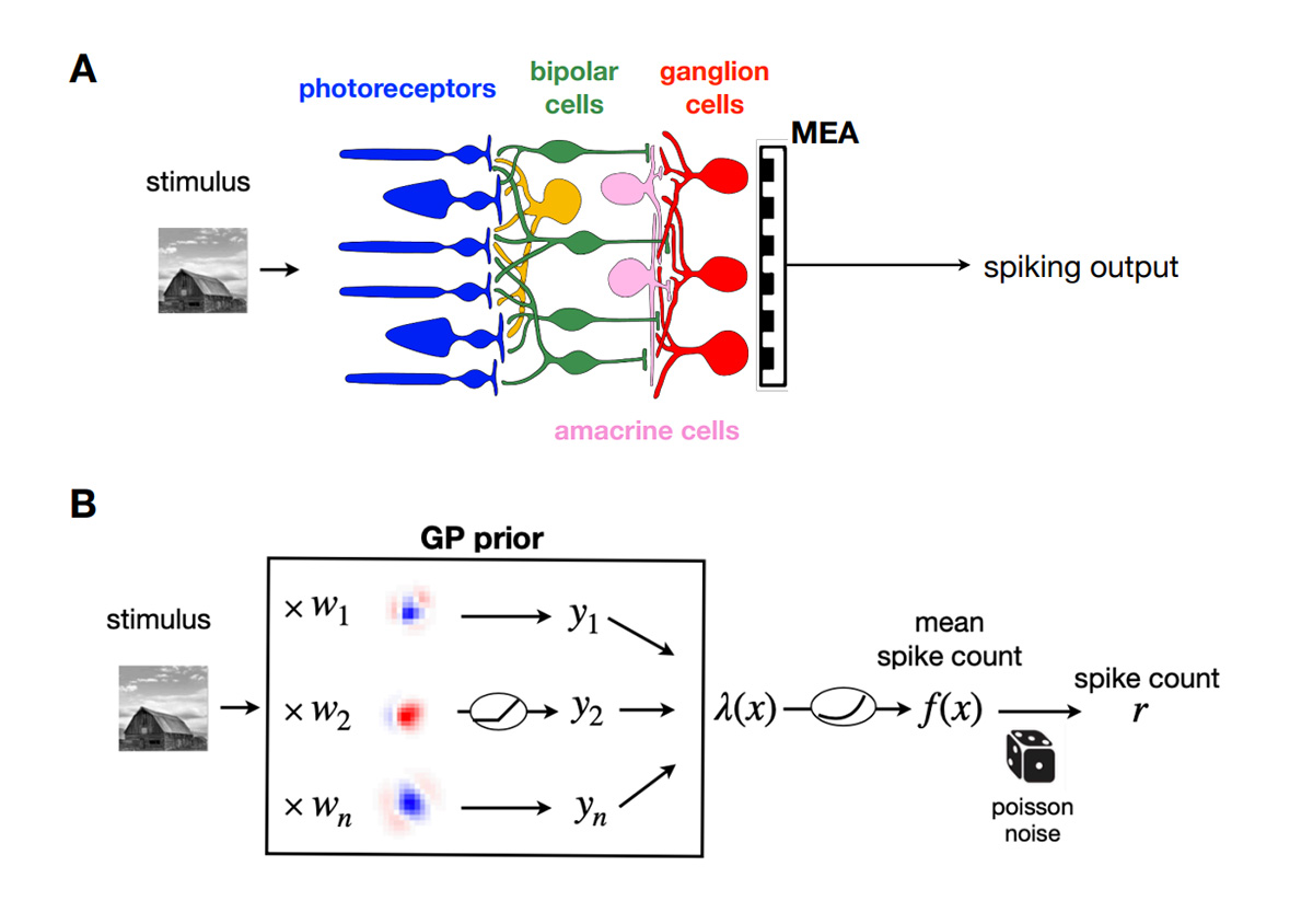
Visual information is processed in the retina. Retinal ganglion cell responses to natural images can be well predicted by a Gaussian process model. The model, schematised here, is equivalent to a 2-layer network.

R1

Based at Sorbonne Université (SU) will focus on developing quantitative models of the function of the retina using a novel optical tool to stimulate individual retinal cells, allowing the differentiation between healthy and degenerated retinas, and other downstream regions, by means of ML algorithms.

START DATE: Month 12

DURATION: 36 months

DELIVERABLES: D1.3, D1.6

R2

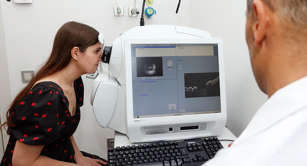
Based at Instituto de Microcirugía Ocular (IMO)  will complement the work carried out by R1 and will focus on the development of new methodologies for the automated clinical evaluation of the visual function based on the precise analysis of ocular movements acquired with eye-tracking technology combined with ML algorithms, to look for patterns that enable early and objective diagnosis of disorders.

START DATE: Month 12

DURATION: 36 months

DELIVERABLES: D1.1, D1.5

OCT analysis of the eye at IMO.
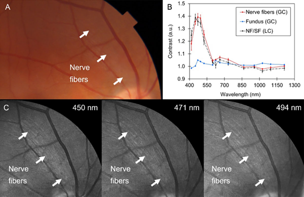
Spectral images of the fundus of the eye and contrast values at different areas.

R3

Based at Universitat Politècnica de Catalunya (UPC)
will extensively exploit MSI together with ML to develop new systems and methods for the analysis of eye structures, retinal oximetry and the automated diagnosis and monitoring of retinal diseases.

START DATE: Month 12

DURATION: 36 months

DELIVERABLES: D1.2D1.7

R4

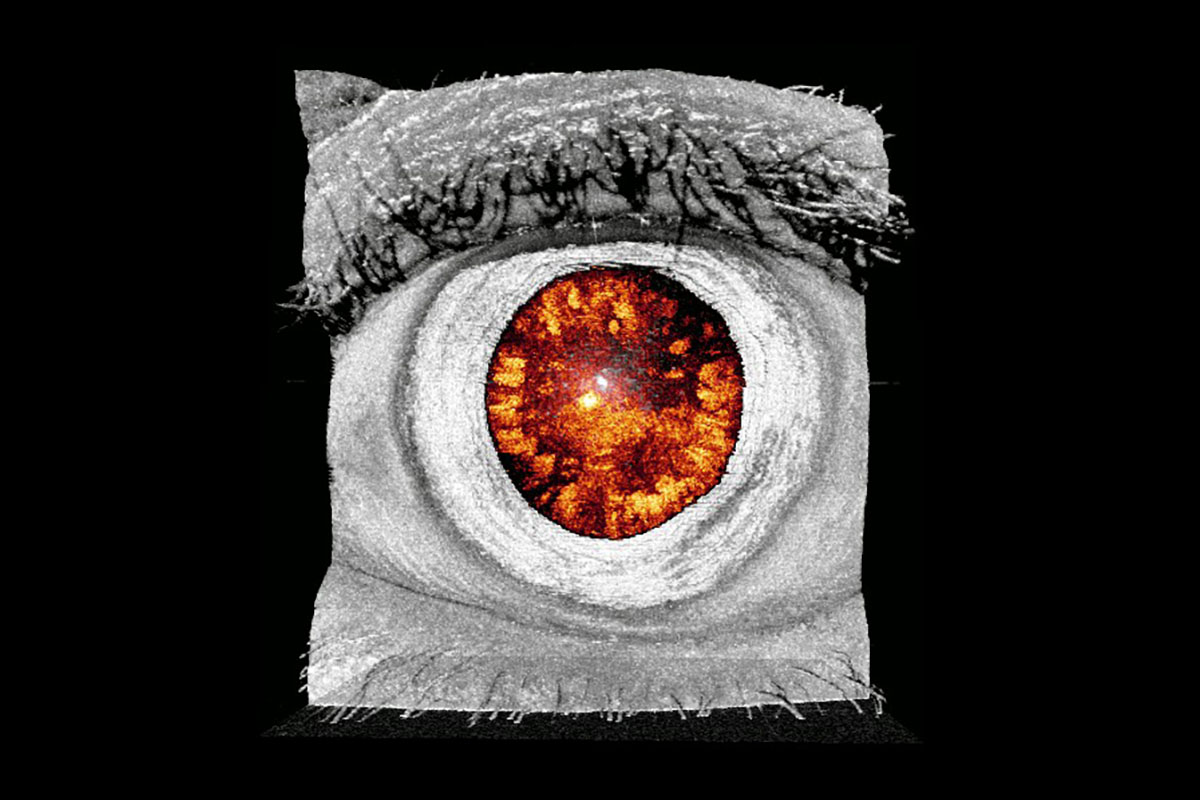
Based at Uniwersytet Mikolaja Kopernika w Toruniu (NCU) will develop a novel tomographic micro-vibrography system for assessing biomechanical properties of ocular structures (cornea, lens, sclera) based on the combination of OCT and optical lock-in detection scheme for high frequency vibrometric measurements. These systems in combination with ML algorithms will be useful to map biomechanical properties of ocular structures.

START DATE: Month 12

DURATION: 36 months

DELIVERABLES: D1.4D1.8

3D reconstruction of a cataract in the eye of a patient.