WP2 – Cardiovascular diseases

“Deaths from cardiovascular diseases surged a 60% globally in the last 30 years.”

The goal of WP2 – Cardiovascular diseases will be to propose new therapeutic approaches to stop the rising incidence of cardiovascular diseases by means of develop and validate optogenetic feedback using advanced optoacoustic imaging and machine learning (ML); by creating ML algorithms to provide early warnings for critical transitions like arrhythmia; also by developing a multimodal imaging tool combined with ML algorithms to analyze facial skin vascularization maps, and in addition, by developing DL algorithms for quantified image acquisition and analysis in clinical multispectral optoacoustic tomography (MSOT), to identify atherosclerotic patients at risk of coronary artery stenosis. In pursuit of this goal, WP2’s research efforts will be conducted across 4 individual research projects in 4 distinct research centers.

Research projects and centers

R5

Max-Planck-Institut für Dynamik und Selbstorganisation (MPI)

SUPERVISOR
Prof. S. Luther (MPI)

CO-SUPERVISOR
Prof. D. Razansky (UZH)

R6

Universitat Politècnica de Catalunya (UPC)

SUPERVISOR
Prof. C. Masoller (UPC)

CO-SUPERVISOR
Prof. U. Parlitz (MPI)

R7

Gdansk University of Technology (PG)

SUPERVISOR
Dr. J. Rumiński (PG)

CO-SUPERVISOR
Prof. J. Pujol (UPC)

R8

Universitat Zurich (UZH)

SUPERVISOR
Prof. D. Razansky (UZH)

CO-SUPERVISOR
Prof. U. Parlitz (MPI)

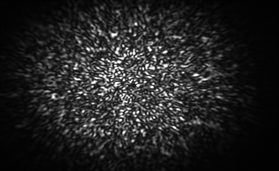
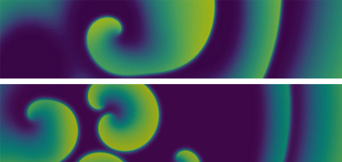
Numerical simulation of rotating spiral waves that resemble the chaotic excitation waves in cardiac arrhythmias.

R5

Based at Max-Planck-Institut für Dynamik und Selbstorganisation (MPI) will develop new methods based on optoacoustic imaging and ML for optogenetic experiments, which can provide new perspectives for optical arrhythmia control. It is important to characterize the electrical waves and calcium concentration in the heart. Using the currently available optical techniques such as a Langendorff perfusion setup with fluorescent dyes, the image quality is still poor and only the activity of the surface of the heart can be monitored. The development of new optoacoustic imaging techniques that will be developed in R5 will seek to eliminate these limitations.

START DATE: Month 12

DURATION: 36 months

DELIVERABLES: D2.3, D2.4, D2.9

R6

Based at Universitat Politècnica de Catalunya (UPC) will develop new algorithms able to provide early warning indication of dynamical transitions to critical states such as arrhythmia and cardiac defibrillation suitable for clinical use. To advance in the clinical application of low-energy anti-fibrillation pacing (LEAP), it is crucial to develop computational models of the heart, which are a powerful way to integrate physical and physiological knowledge in data analysis by ML methods, useful for diagnosis, prognosis and therapy planning.

START DATE: Month 12

DURATION: 36 months

DELIVERABLES: D2.7, D2.8

Speckle image generated by semiconductor light propagating in a multimode fiber. By using image analysis techniques, statistical properties of the light or medium that generated the speckle can be inferred.
Thermal signatures of a face. Respiratory parameters and blood perfusion information can be obtained by observing local temperature changes in time.

R7

Based at Gdansk University of Technology (PG) will focus on advancing the state-of-the-art of multimodal imaging systems, analysing reflected light changes caused by pulsatility of blood in skin facial arteries and thermal imaging. Pulse rate, blood flow and vascularization maps are critical for diagnosis of cardiovascular diseases, in addition to robust ML algorithms for the identification of anormalities in the maps.

START DATE: Month 12

DURATION: 36 months

DELIVERABLES: D2.1D2.6

R8

Based at Universitat Zurich (UZH) will further develop MSOT using image reconstruction methodologies and DL methods. Super-resolution and deblurring machine learning algorithms will be developed to obtain reliable estimations of vascularization maps, which are critical for diagnosis. The goal pursued will be to identify carotid artery stenosis resulting from a build-up of atherosclerotic plaques, which is a major risk factor for ischemic stroke.

START DATE: Month 12

DURATION: 36 months

DELIVERABLES: D2.2D2.5

Multispectral optoacoustic tomography (MSOT) for hand-held, non-invasive diagnostics of carotid artery disease. The image on the right shows the carotid artery bifurcation area acquired with MSOT.