WP3 – Advanced microscopy for the analysis of cells and tissues involved in the development of diseases

“Spatial resolution is now beyond the diffraction limit, enabling the resolution of structural details in cells and tissues.”

The goal of WP3 –Advanced microscopy for the analysis of cells and tissues involved in the development of diseases is to apply new techniques of super resolution microscopy (and try to enhance their capabilities), such as Stimulated Emission Depletion microscopy (STED), Super-resolution Optical Fluctuation Imaging (SOFI) microscopy and One-step Nanoscale Expansion (ONE) microscopy to analyze cells and molecular components in different conditions, such as Parkinson’s disease and muscular dystrophies. In pursuit of this goal, WP3’s research efforts will be conducted across 3 individual research projects in 3 distinct research centers.

Research projects and centers

R9

Georg-August-Universitat Gottingen Stiftung Offentlichen Rechts (UGOE)

SUPERVISOR
Prof. J. Enderlein (UGOE)

CO-SUPERVISOR
Dr. J. Rumiński (PG)

R10

Universitaetsmedizin Goettingen - Georg-August-Universitaet Goettingen - Stiftung Oeffentlichen Rechts (UMG)

SUPERVISOR
Prof. S. Rizzoli (UMG)

CO-SUPERVISOR
Prof. J. Enderlein (UGOE)

R11

Fundació Sant Joan de Déu (FSJD)

SUPERVISOR
Dr. M. Roldán (FSJD)

CO-SUPERVISOR
Prof. M. Vilaseca (UPC)

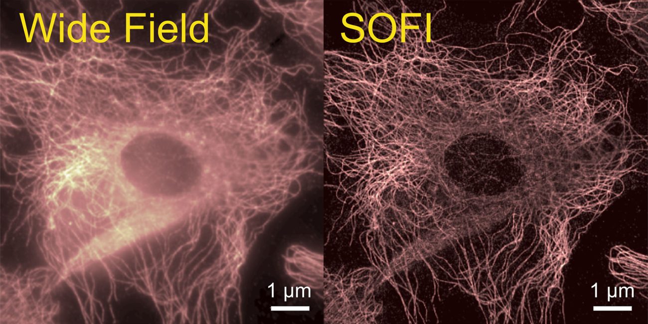
Fluorescently labeled tubulin network in a fixed HELA cell. The left panel is conventional wide field microscopy, the right panel super-resolved SOFI microscopy.

R9

Based at Georg-August-Universitat Gottingen Stiftung Offentlichen Rechts (UGOE) will center its efforts on pioneering novel approaches designed to enhance SOFI’s capabilities with regard to speed, spatial resolution, and the reconstruction of 3D samples.

START DATE: Month 12

DURATION: 36 months

DELIVERABLES: D3.1, D3.4

R10

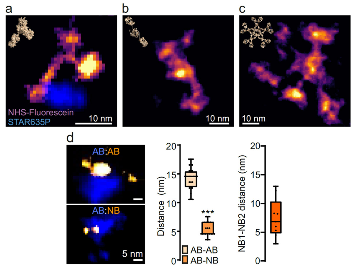
Based at Universitaetsmedizin Goettingen – Georg-August-Universitaet Goettingen – Stiftung Oeffentlichen Rechts (UMG) will apply ONE microscopy, which has spatial resolutions down to 1 nm, and machine learning (ML) for the automated analysis of the shape of single molecules and molecular complexes. This approach holds considerable promise for novel diagnostic applications, particularly in the context of neurodegenerative conditions like Parkinson’s disease.

START DATE: Month 12

DURATION: 36 months

DELIVERABLES: D3.2, D3.5

Images a to c of isolated immunoglobulins (IgG, IgA, IgM), labelled with NHS-ester fluorescein. Distances between fluorescently-conjugated IgGs and fluorescently-conjugated secondary antibodies (top) or secondary nanobodies (bottom).
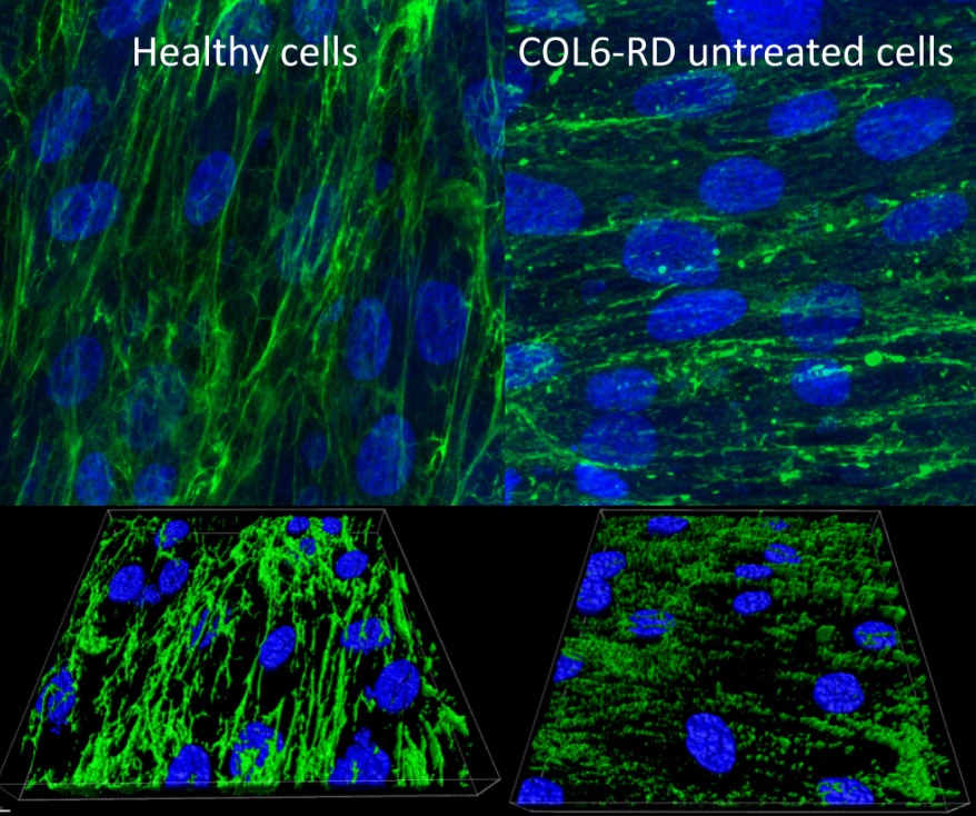
Computational super-resolution images and 3D reconstructions of collagen VI in the fibroblasts from a healthy donor (left), a COL6-RD patient (right).

R11

Based at Fundació Sant Joan de Déu (FSJD) will provide novel methods based on super-resolution microscopy STED and deep learning (DL) algorithms that can aid diagnosis of genetic and acquired rare diseases such as muscular and neurogenetic.

START DATE: Month 12

DURATION: 36 months

DELIVERABLES: D3.2D3.5